Conditions and Services
At Gulf Imaging Associates, we are a team of six fellowship-trained subspecialists, each with unmatched clinical expertise. Our commitment to revolutionizing radiology is evident through our specialized reads, custom-tailored reports, state-of-the-art imaging technologies, quick turnaround times and continuous 24/7 coverage.
Services
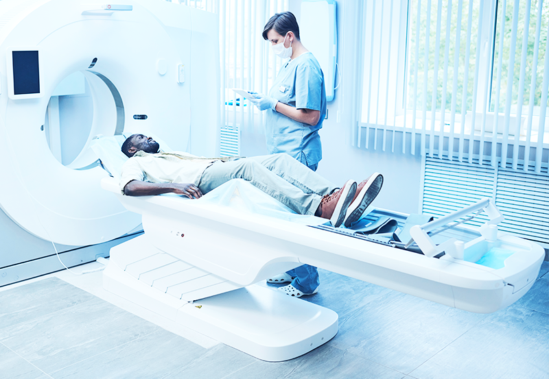
A computerized tomography (CT) scan helps physicians see detailed images inside the body. A CT scan is like taking an X-ray from different angles but rotates around the body, creating cross-sectional images that show more than just bones. A computer then combines the images to create a 3D image to help doctors better understand what is happening inside the body, allowing them to diagnose and treat various injuries, infections and diseases.
Positron Emission Tomography – Computed Tomography (PET-CT) is a unique medical imaging technique combining two types of scans to help doctors see inside the body in greater detail. This technique allows doctors to see the structure of a person’s organs and tissues and how they function. It’s like having two cameras working together to create a complete picture. The PET part of PET-CT uses a special radioactive material called a tracer, which is injected into the body. This tracer flows to different organs and tissues, emitting tiny particles called positrons. The PET scanner can detect these positrons and create a map of where they are in the body. The CT part of PET-CT uses X-rays to create detailed cross-sectional images of your body. By combining PET and CT scans into one, PET-CT provides physicians with more information to diagnose and monitor various conditions.
Magnetic Resonance Imaging (MRI) is a special medical test that uses a strong magnet and radio waves to create detailed pictures of the inside of the body. During an MRI, a patient will lie on a table that slides into a small tube-like machine. Inside the machine, a powerful magnet creates a strong magnetic field around the patient. This magnetic field affects the hydrogen atoms in the body, aligning them in a certain way. Radio waves are sent into the body, causing the aligned hydrogen atoms to produce signals. These signals are picked up by the MRI machine and turned into pictures on a computer. These pictures can show soft tissues like muscles, organs and the brain. MRI scans are completely painless but patients must lie still for the images to appear clear. The entire process typically takes 30 to 60 minutes. During that time, patients can listen to music or wear special headphones to block out the machine noise.
Doctors use MRI scans to help diagnose different medical conditions such as injuries, diseases or organ abnormalities. They are particularly useful for looking at the brain, joints, spine and other parts of the body that cannot be easily seen with other imaging techniques. MRI is a powerful medical tool, helping doctors understand what is happening inside the body and making informed decisions about the best treatment for their patients.
An X-ray machine sends out special beams of light that we cannot see but pass through the body to create images of our bones and other solid structures within the body.
When patients get an X-ray, they might wear a special gown and stand, sit or lie on a table near the X-ray machine. Sometimes, patients may wear a lead apron to protect certain body parts such as the stomach or reproductive organs from radiation exposure. The X-ray technician will give instructions on where to position a patient’s body or parts of the body while they take the picture. Patients do not feel anything but may need to stay still for a few seconds. After the X-rays are complete, patients may get the chance to see the images on a computer screen or printed on a particular film.
Physicians use X-rays to help identify broken bones, infections or problems with a patient’s lungs or other organs.
An ultrasound is a medical test that uses sound waves to create pictures of the inside of the body. Ultrasound is like taking a video or picture using sound instead of light.
During an ultrasound, a special device called a transducer is used. The transducer sends high-frequency sound waves too high for patients to hear. These sound waves travel through the body and bounce back when they hit various parts of the body. The transducer then picks up those bouncing waves and sends them to a computer, turning them into pictures that technicians can see on a screen. These pictures show different parts of our body, such as organs or a baby in the womb. Patients who receive an ultrasound might have to wear a special gown and lie on a table. The technician will put a cool gel on the skin, which helps the sound waves travel better. The technician will move the transducer over the area being examined, pressing it gently against the skin.
Ultrasounds are commonly used to check patients’ organs for problems or abnormalities. It’s a safe and painless process.
Noninvasive vascular imaging allows technicians to look at the blood vessels inside the body without surgery or an invasive procedure. It uses special imaging techniques to create pictures of the blood vessels and observe how the blood flows through them.
One standard method of noninvasive vascular imaging is called ultrasound. Ultrasound uses sound waves to create pictures of our blood vessels. An ultrasound technician will apply a special gel on the skin and gently move a transducer device over the examined area. The transducer sends sound waves into our body and picks up the echoes that bounce back. These echoes are turned into pictures on a computer screen, allowing physicians to see potential problems or blockages.
Other noninvasive vascular imaging methods include magnetic resonance angiography (MRA) and computed tomography angiography (CTA). These methods use powerful magnets and X-rays to create detailed images of the blood vessels. These images help detect blockages, narrowing or abnormalities in the blood vessels that may affect blood flow. Noninvasive vascular imaging is important because it helps physicians identify potential issues in the blood vessels early on. By examining the images created during these procedures, physicians can diagnose conditions like atherosclerosis (a buildup of plaque in the blood vessels), blood clots or aneurysms (abnormal bulging of blood vessel walls). This helps them develop the appropriate treatment plan to improve their vascular health and prevent potential complications like heart attacks or strokes.
Conditions
THE SERVICES LISTED ON THIS WEBSITE ARE FOR GENERAL INFORMATION PURPOSES ONLY AND DO NOT INCLUDE ALL SERVICES OF GULF IMAGING ASSOCIATES. WHILE WE STRIVE TO KEEP THE INFORMATION UP TO DATE AND CORRECT, WE MAKE NO REPRESENTATIONS OR WARRANTIES OF ANY KIND, EXPRESS OR IMPLIED, ABOUT THE CONTENT, COMPLETENESS, ACCURACY, RELIABILITY, LEGALITY, SUITABILITY OR AVAILABILITY, WITH RESPECT TO THE SERVICES CONTAINED ON THIS WEBSITE.